Home

elfogadható Nyomtatás Töltött ultrasound bpd hc ac fl Tanterem Határozza fordulj meg

OB Images
OB Images

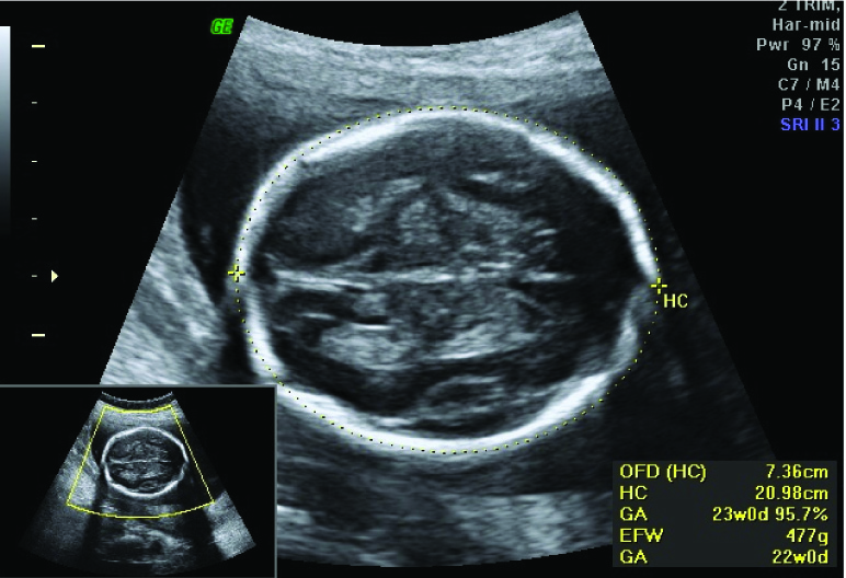
Shown the fetal biometric BPD, HC, AC and FL[Internet web site- | Download  Scientific Diagram
Shown the fetal biometric BPD, HC, AC and FL[Internet web site- | Download Scientific Diagram

shorts BPD,FL,HC,AC parameter measurements (mm)of baby (12 to 40)week by  week in pregnancy - YouTube
shorts BPD,FL,HC,AC parameter measurements (mm)of baby (12 to 40)week by week in pregnancy - YouTube

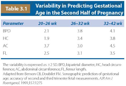
Ultrasound Reference Tables - Williams Manual of Pregnancy Complications,  23 ed.
Ultrasound Reference Tables - Williams Manual of Pregnancy Complications, 23 ed.

Top OB U/S apps: Obstetric Ultrasound Scan Protocols, Fetal Calculator, POC  Ultrasound Guide, Ultrasound Exam Protocols and Image Reference Handbook,  and Ultrasoundpaedia iphone and ipad medical app review
Top OB U/S apps: Obstetric Ultrasound Scan Protocols, Fetal Calculator, POC Ultrasound Guide, Ultrasound Exam Protocols and Image Reference Handbook, and Ultrasoundpaedia iphone and ipad medical app review

jcdr-9-TC01-g004.jpg

Growth, Doppler, and Fetal Assessment | Radiology Key

Fetal Biometry | Obgyn Key

Schematic picture of fetus with HC, AC, FL. HC= head circumference, AC=...  | Download Scientific Diagram
Schematic picture of fetus with HC, AC, FL. HC= head circumference, AC=... | Download Scientific Diagram

How To: Pregnancy BPD HC AC and FL Measurements 3D Video - YouTube

Sonographic Assessment of Fetal Growth Abnormalities | RadioGraphics

Biparietal diameter | Radiology Reference Article | Radiopaedia.org

OB Images
OB Images

Ultrasound scan—fetal growth scan - Patient Information Brochures - Mater  Group
Ultrasound scan—fetal growth scan - Patient Information Brochures - Mater Group

Sex‐specific antenatal reference growth charts for uncomplicated singleton  pregnancies at 15–40 weeks of gestation - Schwärzler - 2004 - Ultrasound in  Obstetrics & Gynecology - Wiley Online Library
Sex‐specific antenatal reference growth charts for uncomplicated singleton pregnancies at 15–40 weeks of gestation - Schwärzler - 2004 - Ultrasound in Obstetrics & Gynecology - Wiley Online Library
BPD, HC, AC and FL values at 28 week scan | BabyCenter

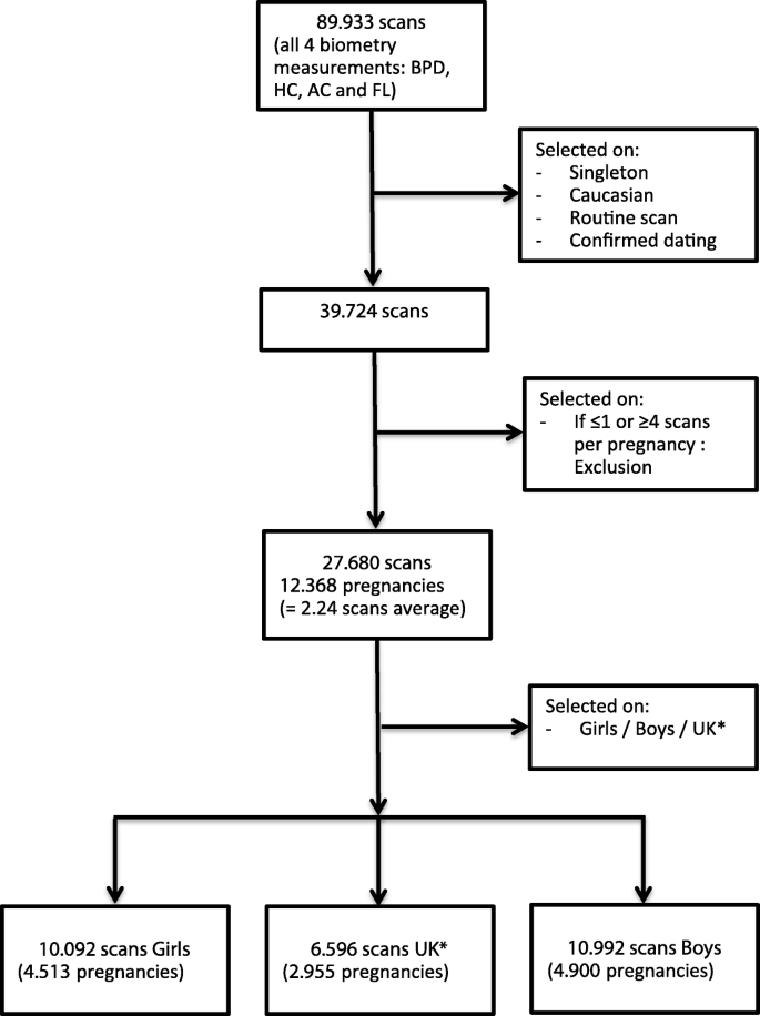
Sex differences in fetal growth and immediate birth outcomes in a low-risk  Caucasian population | Biology of Sex Differences | Full Text
Sex differences in fetal growth and immediate birth outcomes in a low-risk Caucasian population | Biology of Sex Differences | Full Text

Fetal biometry by an inexperienced operator using two‐ and  three‐dimensional ultrasound - Yang - 2010 - Ultrasound in Obstetrics &  Gynecology - Wiley Online Library
Fetal biometry by an inexperienced operator using two‐ and three‐dimensional ultrasound - Yang - 2010 - Ultrasound in Obstetrics & Gynecology - Wiley Online Library

Fetal biometry: Relevance in obstetrical practice - ScienceDirect

Prediction of small-for-gestational age by fetal growth rate according to  gestational age | PLOS ONE
Prediction of small-for-gestational age by fetal growth rate according to gestational age | PLOS ONE

314: Femur length ratios as predictors of adverse outcome in fetuses:  Results from the PORTO Study - American Journal of Obstetrics & Gynecology
314: Femur length ratios as predictors of adverse outcome in fetuses: Results from the PORTO Study - American Journal of Obstetrics & Gynecology

Figure 5 from Detection and Measurement of Fetal Anatomies from Ultrasound  Images using a Constrained Probabilistic Boosting Tree | Semantic Scholar
Figure 5 from Detection and Measurement of Fetal Anatomies from Ultrasound Images using a Constrained Probabilistic Boosting Tree | Semantic Scholar

Normal fetal biometry of a 16 weeks' gestational age | Radiology Case |  Radiopaedia.org
Normal fetal biometry of a 16 weeks' gestational age | Radiology Case | Radiopaedia.org